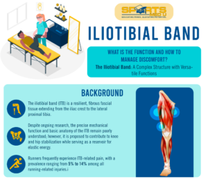
Iliotibial band: What is the function and how to manage discomfort?

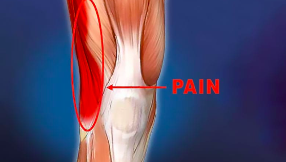
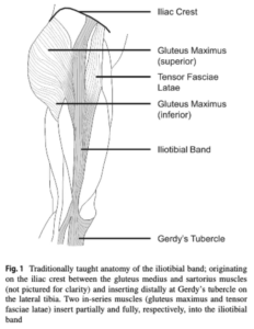
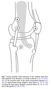
The iliotibial band (ITB) stands as a resilient and fibrous fascial tissue, stretching from the iliac crest to the lateral proximal tibia, intricately woven into human posture when standing.

Despite its pivotal role, the mechanical functions and foundational anatomy of the ITB still pose challenges for full understanding, fuelling ongoing research. Functionally, the ITB has a dual role in stabilising the hip and knee, especially in the frontal plane. Additionally, it’s thought to have potential involvement in storing elastic energy during walking.

However, this seemingly indispensable structure isn’t exempt from issues. Runners often grapple with ITB pain, with a prevalence ranging from 5% to 14% of all running-related injuries. This articles will discuss everything we know so far about the iliotibial band, its function and dysfunction, highlighting key diagnostic tests and the management of iliotibial band syndome.

It will highlight key information from the research study:

Hutchinson, L.A., Lichtwark, G.A., Willy, R.W. and Kelly, L.A., 2022. The iliotibial band: a complex structure with versatile functions. Sports Medicine52(5), pp.995-1008

Join our newsletter and fun infographics that analyse complex sports medicine ressearch

ITB infographic

Anatomical variance

Human Uniqueness in the Iliotibial Band

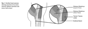
Human anatomy boasts evolutionary uniqueness, especially in the context of the ITB and associated muscles. One standout feature is the human gluteus maximus muscle, which surpasses its non-human primate counterparts in size. This size discrepancy isn’t just a quirk; it plays a pivotal role in enhancing trunk stabilisation, a key adaptation for human erect posture.

Unlike other primates, humans exhibit a structurally distinct ITB, setting them apart anatomically. The human ITB deviates from the fascia lata seen in other primate species, marking a unique evolutionary trajectory.

An additional anomaly lies in the location of the tensor fasciae latae (TFL) muscle in humans. Unlike other animals, the TFL in humans reaches the superior thigh, inserting into the femur near the greater trochanter.

The evolution from walking on all fours to just two is attributed to a combination of factors, including the well-developed gluteus maximus, a shift in pelvic position from horizontal to vertical, and the distinctive formation of the ITB.


Hip muscles


Glutes maximus, tensor fascia lata & the Iliotibial Band

ITB is closely tied to the interplay of the gluteus maximus and TFL. These muscles, with direct insertions, either partial or complete, into the ITB, emerge as contributors to the functional mechanics of this fibrous structure.

In the orchestration of hip movement, the TFL exerts an anterosuperior pull on the ITB, contributing to hip flexion, while the gluteus maximus pulls posteriorly, orchestrating hip extension.

Fascia, a key player in this symphony, is theorised to play a role in broadening muscle insertions by redistributing or redirecting force transmission within the musculature. The ITB, by broadening the insertion of the gluteus maximus muscle and facilitating TFL muscle insertion, emerges as a central figure in transmitting forces from these muscles across both the knee and hip joints.

However, despite these findings, the specific function of the ITB and the nuanced impact of variations in activation levels of the muscles remain largely unknown. Understanding this biomechanical puzzle holds promise for a deeper understanding of human movement, providing valuable insights for athletes, clinicians, and researchers alike.

Variability in Iliotibial Band Insertions and Implications for Function

The distal insertion of the ITB stands as the most intriguing mystery, with descriptions varying widely among researchers. While everyone agrees that the ITB inserts at Gerdy’s tubercle, the literature presents a diverse array of alternative distal insertion points. These include:

  • The insertion point of the lateral femorotibial ligament
  • Supracondylar femur (Kaplan’s distal fibers)
  • Along the linea aspera (Kaplan’s proximal fibers)
  • Patella.

Recent literature introduces a paradigm shift, challenging the conventional notion of the ITB as a distinct structure. Instead, it suggests that the ITB might be essentially a thickening of the fascia lata.

The inconsistencies in the literature regarding the distal insertions of the ITB pose challenges in understanding its function fully. However, it prompts some intresting questions – do these diverse insertions indicate distinct force transmission pathways within the ITB? Could this variability reflect its numerous potential mechanical functions, influenced by factors such as posture and muscular activation? As we go through the article, the varied descriptions may unveil the nuanced adaptability and complexity within the ITB’s role in human biomechanics.


ITB possible insertions

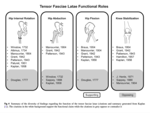
Tensor Fascia Latae Biomechanics

The consensus supports TFL’s involvement in hip internal rotation, hip flexion, and knee stabilisation, however its high electromyographic (EMG) activity during isolated abduction has fuelled debates surrounding its primary function.

Speculation regarding TFL’s potential role in hip abduction exists, but the prevailing view contends that any force it exerts at the knee is transmitted via the ITB, implying a stabilising role at the knee rather than an active contributor to joint movement.

Gottschalk et al.’s innovative concept positions the ITB as a “strut” during normal walking, providing frontal plane stabilisation for the hip. Recent research by Neumann  delves into the theoretical potential actions of hip muscles, suggesting that TFL, with its frontal plane moment arm, may play a vital role in stabilising the pelvis in the frontal plane.

Notably, the mechanical role of TFL likely hinges on the posture of the ITB during force production, considering its double joint nature and shared insertion onto the ITB with the gluteus maximus, a muscle opposing hip flexion.

TFL’s biomechanics not only highlights the complexities within the hip musculature but also emphasises the interconnected nature of muscles and their roles in maintaining stability and orchestrating movement throughout the body.

TFL attachments


Gluteus Maximus: A power house with Unanswered Questions

The gluteus maximus, hailed as a primary hip joint extensor, stands as a muscular powerhouse with remarkable attributes. Its substantial muscle volume and a significant hip extension moment arm in the sagittal plane emphasises its  role in powerful movements. Beyond its primary role in hip extension, gluteus maximus is a versatile player, contributing to external hip joint rotation, hip abduction, and notably, the tensioning of the iliotibial band.

Given a substantial proportion of its fibers inserting onto the ITB, gluteus maximus likely plays a big role in transmitting greater force through the ITB compared to the TFL.

Despite its biomechanical significance, the portion of the gluteus maximus muscle that inserts on the ITB remains a subject of considerable debate. Gluteus maximus, with its different muscular jobs and unanswered questions, stands as both a biomechanical marvel and a subject of ongoing exploration in the scientific world.

Gluteus maximus ITB insertion

Knee Stability: Insights into ITB Dynamics

Gluteus maximus and TFL insertions into the ITB has become a focal point for researchers exploring their potential impact on lateral knee stabilisation. Particularly, the attachments to the patella emerge as a key consideration, offering a stabilising mechanism against medial dislocation and contributing to patellar stability.

The ITB’s connections to the anterior (front) and lateral (outside) tibia form a critical line of defence against anterolateral subluxation, a mechanism crucial in the pivot shift of an anterior cruciate ligament ACL-deficient knee. However, the  knee stabilisation mechanisms involving the ITB are varied, influenced by factors such as attachment location, loading, mechanical behaviour, and posture.

While ITB insertions alone may not  prevent anterior dislocation in a fully extended knee with ACL deficiency, research highlights their capacity to reduce anterior translation beyond 30° of knee flexion, particularly notable in the pivot shift mechanism.

It is essential to note that much of this research is grounded in cadaveric studies, presenting a limitation as these studies predominantly involve passive forces, overlooking the potential substantial contributions from dynamic activities like walking and running, where gluteus maximus, and TFL may exert significant forces.

The quest for a precise understanding of the ITB’s contribution to knee stability persists, requiring a deeper exploration of how forces are applied in diverse postures and dynamic scenarios.

ITB distal attachments

The impact of ITB on compression forces

ITB’s resistance to external adduction type forces after direct impact and the knee compression force to the femur through ITB tensioning contribute significantly to achieving stability.

Magnetic resonance images provide a visual testament to the impact of ITB tensioning, showing compression in the tissues between the distal ITB and the lateral femoral epicondyle. This compression mechanism, integral to knee joint stability, demonstrates the interesting dynamics at play during movement.

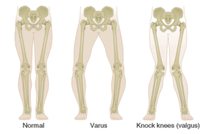
However, the delicate balance between stability and potential challenges comes to the forefront in discussions surrounding iliotibial band syndrome (ITBS). Current theories propose ITBS as a compression syndrome, suggesting that the enhanced knee joint stability provided by the ITB may, in some instances, lead to potentially unfavourable excessive compression. This is particularly notable in the presence of varus knee torques, where the forces on the knee are directed inward, potentially exacerbating compression-related issues.

Varus knee

In lab & out of lab material properties and elastic function

Material Properties of ITB & understanding how it affects biomechanics

The material behaviour of the ITB is pivotal to know if we want to understand the roles it plays in human movement and stability. Acting as a vital connection point for muscles, such as the gluteus maximus and TFL, to bones like the pelvis, femur, and tibia, the ITB suggests tendon-like material properties, contributing significantly to joint stability and potentially participating in elastic energy storage and release, just like the Achilles tendon.

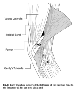
While the general material properties of the ITB are well-documented, the overall mechanical behaviour of the entire structure remains somewhat blurred. Tensioning of ITB fibers during hip extension follows an anterior-to-posterior pattern, resulting in varied tension across different regions of the band based on movement patterns.

The transmission of forces within the ITB unfolds as a diverse process, influenced by the specific muscles generating forces.

Elastic Function of the ITB in Human movement

The human lower limbs are marvels of biomechanical efficiency, featuring spring-like tendons that optimise the economical storage and release of energy during movement. Among these energy-saving structures are the well-known Achilles tendon, plantar fascia, ITB, and peroneus longus.

The Achilles tendon takes the lead, making a substantial contribution of around 35-40% (35 J) of work during the stance phase of running. Recent research has shown that the ITB has a major role in energy storage & release, suggesting it can store up to 5% of the total work in a moderately paced run, roughly 14% of the work contributed by the Achilles tendon.

Eng et al. model adds another layer to our understanding, proposing that the posterior ITB, particularly the portion with the gluteus maximus muscle insertion, can transmit larger forces than the anterior portion with TFL muscle insertion. This asymmetry potentially leads to greater energy absorption.

Despite these insights, assessing the energetic contributions of soft tissues like the ITB remains challenging. The numerous degrees of freedom and the difficulties in directly accessing their kinetic contributions to motion add complexity to the study of these spring-like structures.

ITB elastic energy when running

Clinical Significance

ITB syndrome –  Is it friction or compression that causes the issue?

Iliotibial Band Syndrome (ITBS) stands out as a prevalent overuse injury with profound implications for athletes engaged in activities like running and cycling. Characterised by lateral knee pain, particularly exacerbated during actions like single-leg stance, ITBS holds the position of being the most common relative overuse injury at the lateral knee, contributing to approximately 12% of all running-related injuries and making substantial impacts in cycling and military-related contexts.

Historically perceived as a friction injury, the conventional wisdom held that cyclic loading during activities like running and cycling led the iliotibial band to rub the lateral epicondyle, causing irritation to the innervated fatty tissue beneath.

However, recent insights by Fairclough et al. have shaken this foundational belief, suggesting that the perceived friction might be an illusion. Their questioning of the friction mechanism proposes an alternative view, emphasising the sequential load shifting of ITB fibers from anterior to posterior during tensioning.

Adding a new dimension to our understanding, a novel theory supported by magnetic resonance imaging (MRI) suggests that when the knee flexes beyond 30°, the ITB compresses medially against the lateral femoral epicondyle. This compression mechanism introduces a paradigm shift, proposing that ITBS should be classified as a compression syndrome rather than a friction injury.

As we delve deeper into the pathomechanics of ITBS, this evolving narrative contributes not only to a refined understanding of the condition but also paves the way for more targeted and effective approaches to its prevention and treatment.

ITB and the fat pad benath

Running Kinematics in Iliotibial Band Syndrome

Running kinematics in individuals developing ITBS reveal movements that distinguish them from healthy controls. Studies by Noehren et al. and Friede et al. observe increased hip adduction and knee internal rotation in those prone to developing ITBS during running.

Contrastingly, runners with prior ITBS history exhibit reduced hip adduction compared to their healthy counterparts. This paradoxical finding suggests that individuals who have experienced ITBS may alter their running patterns as a protective adaptation.

Furthermore, investigations show a gradual decline in peak hip adduction angle during prolonged runs in individuals with ITBS. This decline may indicate a strategic shift in running kinematics, possibly adopted to alleviate pain or mitigate strain on the ITB as the run progresses.

In essence, these collective findings suggest that runners, whether prone to or with a history of ITBS, may adopt altered movement patterns in response to pain or injury, showcasing the remarkable adaptability of the human body.

Diagnosis and Differential Considerations

Diagnosing ITBS involves a keen understanding of the distinctive symptoms and potential differential diagnoses. Runners grappling with ITBS commonly report lateral knee pain, specifically situated around 2–3 cm proximal to the lateral tibiofemoral joint line, particularly emphasising the lateral femoral condyle.

The onset of ITBS pain typically follows an insidious pattern, often linked to recent surges in running loads. This surge might manifest as an increase in running distance or a higher volume of downhill running, signalling a critical clue for diagnosis.

Individuals with particularly irritable ITBS may experience pain during stair descent in the stance limb, especially during hip extension and knee flexion. This discomfort aligns with the eccentric contraction of the TFL muscle, contributing to lower limb control.

For an accurate diagnosis, clinicians must rule out alternative sources of lateral knee pain. These may include:

  1. Patellofemoral pain
  2. Lateral meniscal lesions
  3. Lateral synovial plica syndrome
  4. Distal femoral bone stress injuries.

Specific clinical assessments and screenings play a crucial role in this process.

Additionally, considering conditions like gluteal tendinopathy and lumbar radiculopathy is important, as these may refer pain to the lateral thigh and knee.

Noble Compression Test: A Diagnostic Tool for ITBS

When it comes to diagnosing suspected Iliotibial Band Syndrome, the Noble compression test takes the lead as a primary diagnostic evaluation. This straightforward yet informative test involves the application of manual pressure to the lateral knee, specifically targeting a point 1–2 cm proximal to the lateral femoral condyle. The knee is then passively extended through a range of motion from 60° to full extension.

A positive result in the Noble compression test is identified when the maneuver elicits the reproduction of lateral knee pain, particularly when the knee is positioned at approximately 30° of knee flexion. This specific angle is crucial for the sensitivity of the test, capturing potential discomfort associated with ITBS.

However, it’s essential to approach test interpretation with caution, as the Noble compression test currently lacks established positive and negative likelihood ratios.

Nobles test for ITBS

Ober’s Test and Hip Weakness in ITBS

The Ober test has become a go-to tool for clinicians assessing ITB “tightness,” with both classic and modified versions widely employed. Despite the common assumption that an injured ITB is inherently tighter than a healthy one, neither iteration of the Ober test appears to effectively measure this perceived “tightness.”

A positive result in the Ober test often indicates restrictions in the hip capsule and the musculature of the gluteus medius and minimus. However, it’s crucial to note that neither version of the Ober’s test is particularly helpful in diagnosing ITBS or assessing ITB “tightness.”

In individuals with ITBS, a prevalent observation is hip abductor weakness. Interestingly, this weakness is considered a consequence of ITBS rather than a contributing factor. The relationship between hip abduction strength and ITBS remains unclear due to the absence of prospective studies. It is hypothesised that pain arising from distal compression within the highly innervated tissues of the ITB may hinder proximal hip musculature.

Adaptive engagement of the TFL and gluteus maximus in strategies to diminish tension in the ITB may further complicate the relationship. Thus, the presence of hip weakness appears to be concurrent with ITBS rather than being a causative factor in the onset of this injury.

Obers test for ITBS

Treatment for ITBS: Progressive Overload and Graded Exposure

The treatment of Iliotibial Band Syndrome is a topic often explored in scientific literature, although the quality of available evidence is frequently constrained to narrative reviews or case series, typically of lower quality.

Despite these limitations, a consistent and widely recommended approach for individuals seeking recovery from ITBS involves the integration of progressive overload and graded exposure to progressively challenging activities. This  strategy acknowledges the need for a gradual increase in intensity, allowing the affected tissues, particularly the iliotibial band, to adapt and strengthen over time.

Progressive overload entails a systematic and incremental increase in the demand placed on the body during physical activity. This may involve modifying running surfaces, distances, or incorporating specific strength and conditioning exercises targeting the hip and knee musculature.

Graded exposure emphasises a gradual re-introduction of activities that may have contributed to or exacerbated ITBS symptoms. This approach allows individuals to acclimate to the demands of running or other activities while minimising the risk of symptom recurrence.

While the literature may lack high-quality evidence, the consensus on the importance of progressive overload and graded exposure suggests a practical and pragmatic foundation for managing ITBS. Individualised treatment plans, incorporating these principles, can empower individuals to regain function and resume their desired level of physical activity with reduced risk of recurrence.


Foam Rolling and Stretching for ITB

Foam rolling of the ITB has become a popular recommendation for runners grappling with perceived ITB “tightness” or ITBS. However, despite its widespread use, the effects of foam rolling on flexibility appear to be short-lived or minimal, with any pain relief achieved being temporary and lasting only a few minutes.

Stretching the ITB, while often suggested, presents challenges, especially in the context of assessing ITB “tightness” as exemplified by the Ober test. This test, commonly used in clinical settings, may not effectively measure true ITB “tightness.”

A recent study by Friede et al. delved into ITB stiffness using shear-wave elastography, investigating both healthy participants and those experiencing ITBS symptoms after a 6-week training period. Surprisingly, the study found no discernible differences in ITB stiffness between healthy individuals and those with ITBS.

These findings show the limitations of common strategies like foam rolling and stretching when addressing ITB-related concerns. While these techniques may offer temporary relief, a more comprehensive approach that considers individualised factors contributing to ITBS and incorporates progressive and graded interventions may be a lot more beneficial.

Hip Strength and ITBS

Hip strengthening has emerged as a common recommendation for individuals grappling with ITBS. While a hip-strengthening program has demonstrated concurrent resolution of hip weakness and pain in runners with ITBS, caution is urged in attributing causation to hip weakness, as it typically follows the onset of ITBS.

Interestingly, hip strength does not appear to correlate with hip adduction during running, challenging a prevalent belief among clinicians. Contrary to expectations, engaging in hip strengthening exercises does not necessarily result in a reduction in hip adduction during running.

An alternative explanation for the pain relief experienced by individuals with ITBS following hip strengthening lies in the potential of targeted loading exercises, like intensive hip strengthening, to alter central pain processing and diminish local hyperalgesia. This suggests that the benefits of hip strengthening may extend beyond solely addressing biomechanical factors, influencing broader pain perception and tolerance.

Running Biomechanics for ITB Health

Addressing running biomechanics holds promise in mitigating suspected increases in ITB strain and subsequent compressive loads on the lateral knee. A study by Meardon et al. utilised a subject-specific musculoskeletal model during running, uncovering that adopting a wider step width effectively reduces ITB strain.

Implementing interventions to optimise running biomechanics can be straightforward and practical. Providing feedback on step width, a key factor in influencing ITB strain, can be easily achieved using a full-length mirror during treadmill running, offering real-time visual cues for runners.

Another impactful intervention involves increasing running cadence, also known as stride frequency. This adjustment has demonstrated the ability to reduce both ITB strain and strain rate, as evidenced by a comparable musculoskeletal model. Encouraging an uptick in running cadence aligns with practicality, as it can be seamlessly implemented during routine, in-field runs through the use of commercially available wearable devices, providing real-time feedback to runners.

Join the sports medicine community and get free break downs of sports medicine journals

Treatment for iliotibial band syndrome

Source:

  • Eng, C.M., Arnold, A.S., Lieberman, D.E. and Biewener, A.A., 2015. The capacity of the human iliotibial band to store elastic energy during running. Journal of biomechanics48(12), pp.3341-3348.
  • Fairclough, J., Hayashi, K., Toumi, H., Lyons, K., Bydder, G., Phillips, N., Best, T.M. and Benjamin, M., 2006. The functional anatomy of the iliotibial band during flexion and extension of the knee: implications for understanding iliotibial band syndrome. Journal of anatomy208(3), pp.309-316.
  • Friede, M.C., Klauser, A., Fink, C. and Csapo, R., 2020. Stiffness of the iliotibial band and associated muscles in runner’s knee: Assessing the effects of physiotherapy through ultrasound shear wave elastography. Physical Therapy in Sport45, pp.126-134.
  • Gottschalk, F.R.A.N.K., Kourosh, S.O.H.R.A.B. and Leveau, B., 1989. The functional anatomy of tensor fasciae latae and gluteus medius and minimus. Journal of anatomy166, p.179.
  • Hutchinson, L.A., Lichtwark, G.A., Willy, R.W. and Kelly, L.A., 2022. The iliotibial band: a complex structure with versatile functions. Sports Medicine52(5), pp.995-1008
  • Meardon, S.A., Campbell, S. and Derrick, T.R., 2012. Step width alters iliotibial band strain during running. Sports Biomechanics11(4), pp.464-472.
  • Neumann, D.A., 2010. Kinesiology of the hip: a focus on muscular actions. Journal of Orthopaedic & Sports Physical Therapy40(2), pp.82-94.

Leave a Reply

Your email address will not be published. Required fields are marked *